 Abstract
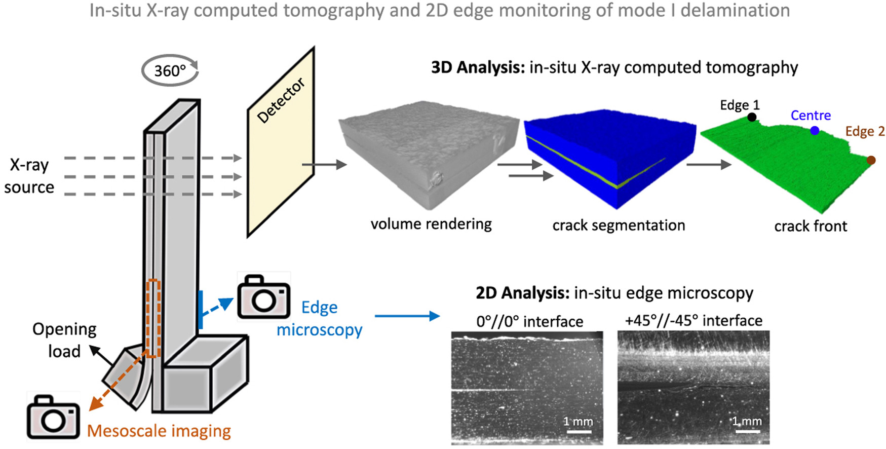
Test methods determining interlaminar fracture toughness of composites are typically performed in conjunction with in-situ surface imaging techniques to detect the crack initiation and monitor its propagation. However, these methods struggle to visualise the 3D evolution of the crack front. In such tests, initiation detection is based on force-displacement curve features, such as the non-linearity point, or edge observations. This paper reports the implementation of a methodology for in-situ 3D characterisation of the crack front in a double cantilever beam test using microfocus X-ray computed tomography. The crack initiation and propagation inside the bulk were determined through crack segmentation in the in-situ tomograms, allowing quantification of the fracture toughness without the need for artificially imposed initiation criteria such as the non-linearity. Two different delamination interfaces were investigated: 0°//0° and +45°//−45°. These two interfaces can be viewed as two extremes in terms of delamination behaviour. The results of the proposed methodology are compared against those measured via the “conventional” edge microscopy coupled with digital image correlation.
Graphical abstract