The KU Leuven XCT Core Facility offers X-ray Computed Tomography (XCT) infrastructure and services to study the internal structure and behavior of materials and biological tissues at different length scales. For X-ray methods the contrast in the images is the result of a mixed combination of density and compositional information. X-ray computed tomography (XCT) therefore allows reconstructing the 3D internal structure of objects in a non-destructive way, without (or with limited) prior sample preparation. The core facility houses in total 7 complementary micro- and nanofocus XCT systems and in-situ testing devices, e.g., loading stages, pressure cells and temperature and moisture controlled cells. For advanced image processing and analysis of the high resolution 3D and 4D images, the core facility disposes of different commercial and non-commercial software packages (Voxtex, Matlab, Avizo, CTAn, …).
To make the KU Leuven XCT core facility (KUL-XCT) accessible for a broad scientific audience and for services to industry. The core facility plays an important role in ground-breaking research to unravel the internal structure, morphology, microstructural changes or damage development in materials, products and biological specimens, and provides the scientists with high quality XCT images from which quantitative data for the fundamental understanding of mechanisms and for modelling can be retrieved.
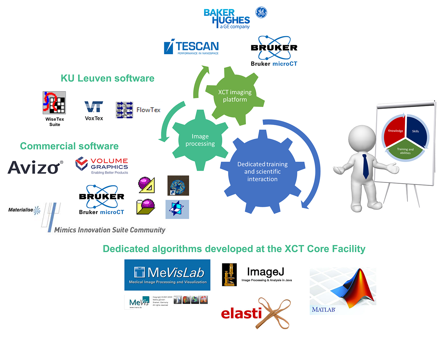
The three major pillars
A multiscale 3D-XCT imaging platform, allowing to asses materials from full sized samples (i.e. organs, fruits, geological cores, micro-electronic chips, building materials, etc.) down to the level of the smallest building blocks (i.e. cells, crystals, grains, micro-electronical components, etc.) and providing improved insight into the relation between the functionality and the 3D morphology.
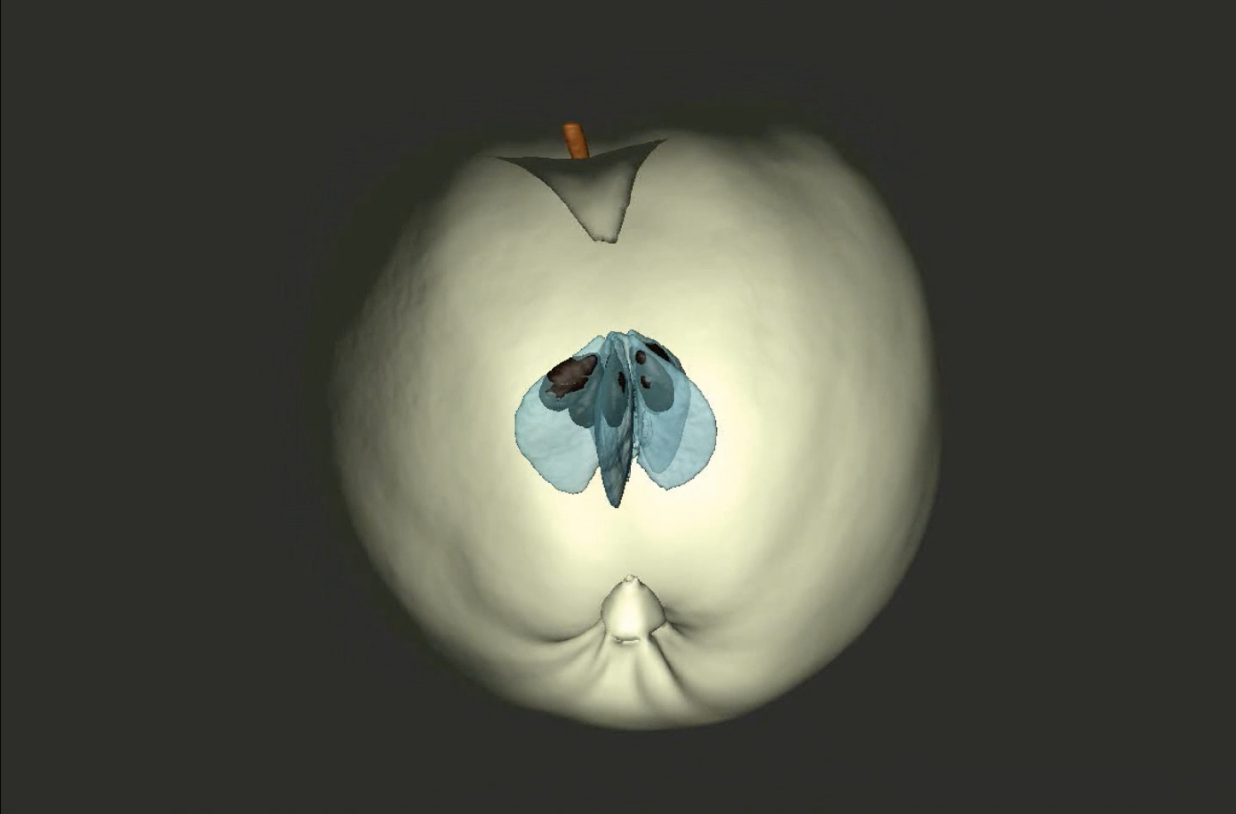
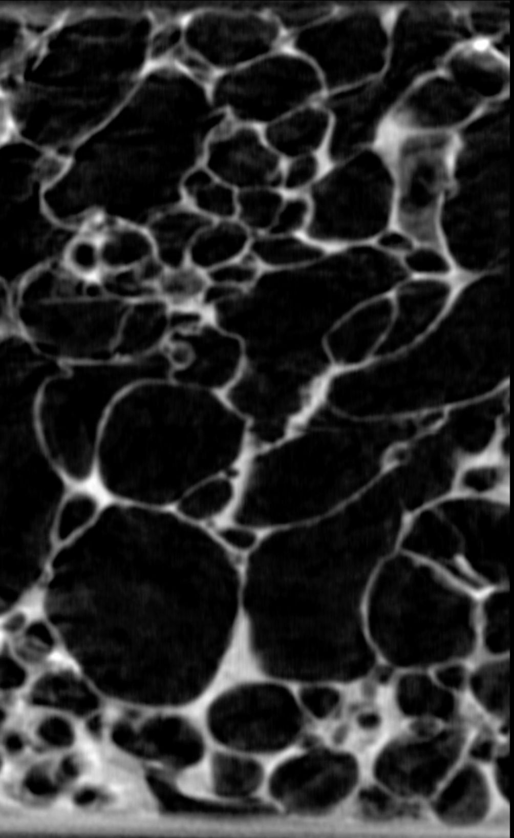
This movie shows the microstructure of an apple and was rendered from (microfocus) CT images of an apple. The images of the entire apple were obtained with an X-ray CT system at KU Leuven (MTM) and the images of the tissue were obtained at the ID19 beamline at the European Synchrotron Radiation Facility at 5 micrometer pixel resolution (Plant Physiology, June 2008, Vol. 147, pp. 518–527). These results were partially funded by the European Commission (project InsideFood).
XCT-scanning in a controlled environment (temperature and moisture), as well as during loading, wetting, etc. (4D-XCT, in which time acts as the 4th dimension).
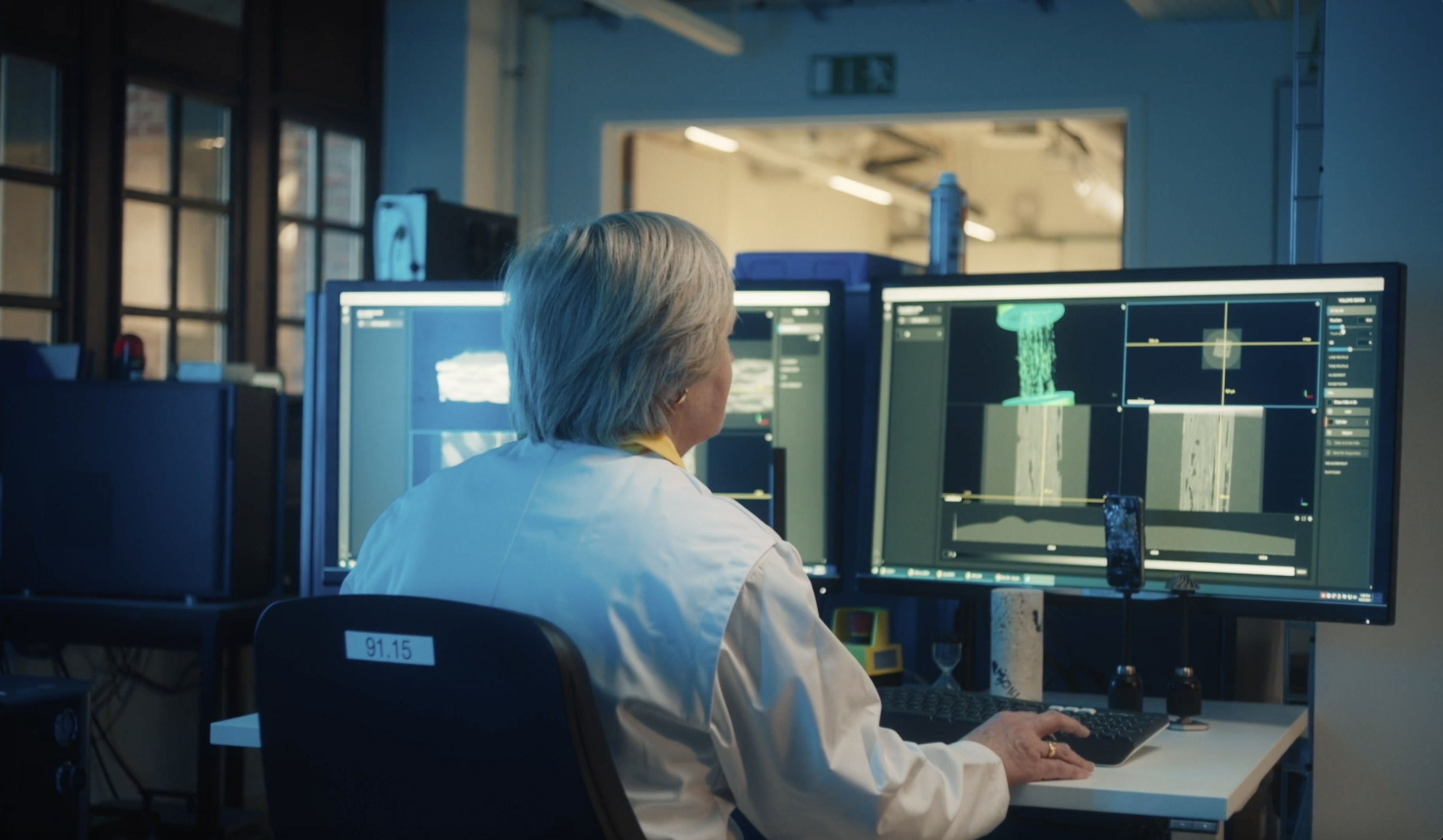
XCT facilities managed interdisciplinary by skilled personnel pushing the equipment to a higher scientific level and providing users with the required technical and scientific support.