
RL4 – BREATHE Lab
MicroCT allows the non-destructive assessment of ex vivo human pulmonary specimens with high resolution. This enables airway and vascular segmentation and quantification to study morphological changes in health and in chronic respiratory disease. Given the non-invasive characterization of the lung, further downstream processing is not limited and allows further use of the specimen for advanced molecular investigation
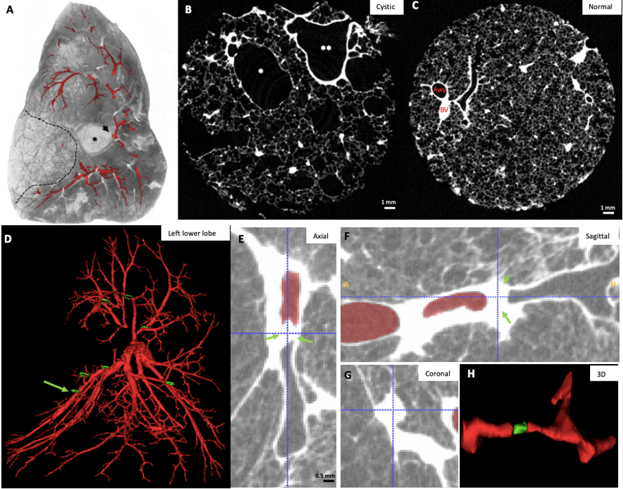
Figure 1. 3D airway morphology in congenital pediatric pulmonary disease. A. Cross-sectional whole lobe microCT image of 3D airway tree segmentation. B. Cross-sectional microCT image in an area of cystic destruction illustrates the cystic destruction (*) C. Cross-sectional microCT image in a non-cystic area within the same lobe demonstrates normal parenchyma and normal conducting airways (Awy) with accompanying blood vessels (BV) D. 3D segmentation of the airway tree (microCT scan) shows abundant airway atresia/obstruction (green circles). One representative airway lesion (green arrow) is further highlighted. E. Axial view of the airway lesion demonstrating complete obstruction of the airway lumen with re-opening of the airway lumen distally from the obstruction without direct connection to the bronchial tree. Sagittal (F) and coronal (G) view of the airway lesion confirm complete airway obstruction. H. 3D reconstruction of this obstructed airway illustrates the blunt ending airway (obstructed segment in green) and distal re-opening and normal appearing bifurcation.