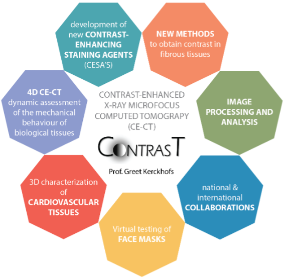
Histology is the gold standard for assessing biological tissues. However, it is a destructive, costly, and time-consuming technique, and is mostly restricted to 2D sections. It, therefore, has limitations with regards to the structural assessment of 3D biological tissues and the quantification of the spatial organization of the tissue constituents, such as collagen and elastin fibres, proteins, etc. X-ray CT provides a solution for mineralized tissues, but soft tissues remain invisible due to their low X-ray attenuation. The ContrasT Team, under the supervision of Prof. Greet Kerckhofs, performs pioneering work with regards to 3D contrast-enhanced microCT (CE-CT) imaging of soft biological tissues. CE-CT can tackle the shortcomings of histology as it allows full 3D soft tissue visualization, and subsequent 3D microstructural analysis. The ContrasT Team has a highly interdisciplinary expertise (biology, chemistry, engineering, imaging), and attempts to develop and validate (i) novel contrast-enhancing staining agents (CESAs) for specific tissues and proteins and (ii) novel contrasting methods, and to apply these to different biological (sub)tissues.
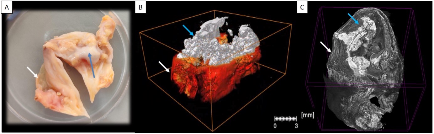
Calcification of human aortic heart valves. (A) A leaflet of a human aortic valve. (B) A microCT-based 3D rendering of a human aortic leaflet, visualizing the calcifications; the soft tissue is visible in red, but there is no information on its fibrous organization. (C) CE-CT-based 3D rendering of the human aortic leaflet, visualizing the calcifications, but also the fibrous organization of the soft tissues. White arrows indicate the soft tissue, blue arrows indicate the calcifications