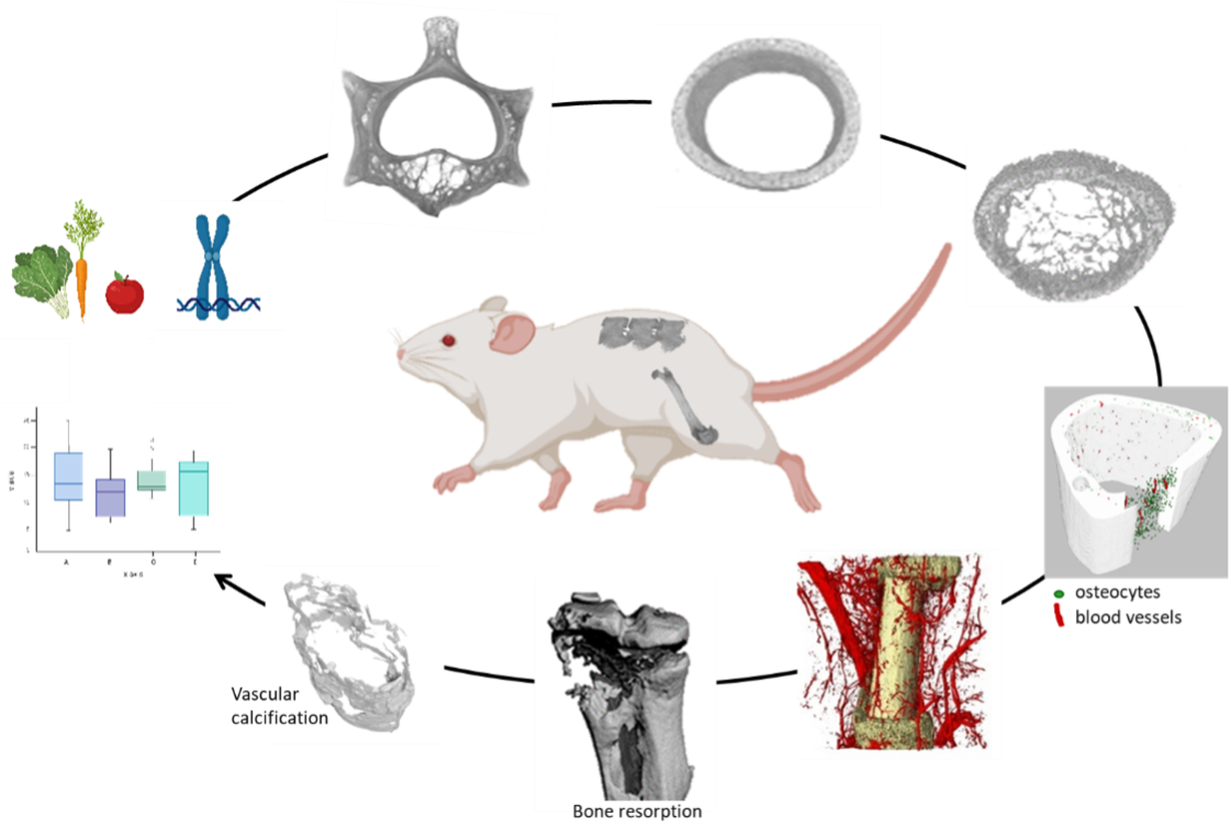
X-ray attenuation methods like XCT can be used to assess the mineralized matrix in biological tissues. Based on image segmentation, 3D quantitative analysis provides more insight in the development, maintenance and repair of mineralized tissues such as bone, but could also improve our understanding of pathologies characterized by altered bone mass (e.g. osteoporosis, osteoarthritis, bone cancer/metastasis) or undesirable mineralization (kidney stones, vascular calcification). Moreover, by using specific contrast agents, dual energy imaging facilitates simultaneous analysis of mineralized and non-mineralized tissues, for example bone and adjacent vasculature. In addition to ex vivo analysis of post-mortem dissected samples (rodent tissues or human biopsies), XCT can also be employed for in vivo analysis of mineralized tissues in living, anesthetized animals. Protocols are optimized to avoid detrimental side effects caused by repetitive X-ray dosing.