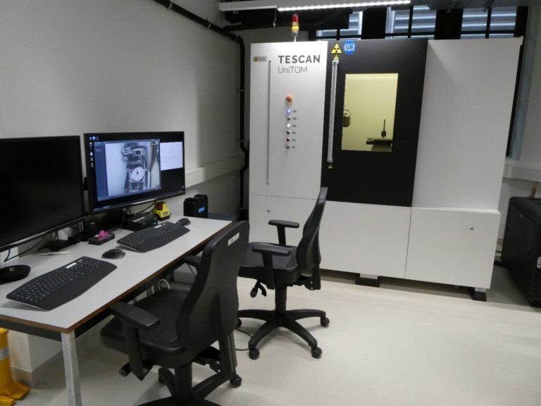
TeScan Unitom HR
The Unitom HR is a modular and flexible sub-micron resolution XCT system. The system aims at making high resolution and high contrast images.
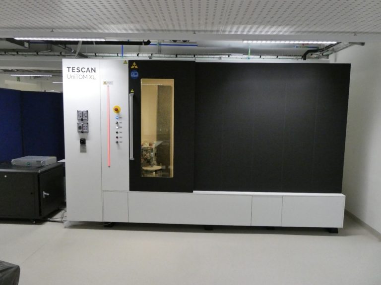
TeScan Unitom XL
The Unitom XL is a multi-resolution micro-CT, optimized for high throughput and contrast, diverse sample types, and flexibility for your research.
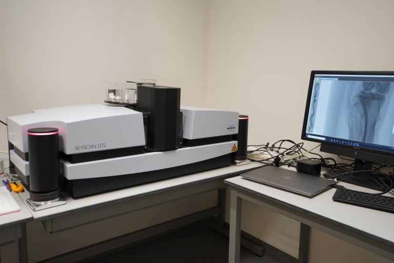
Bruker SkyScan 1272
The Skyscan 1272 is a high-resolution desktop X-ray microtomograph, optimized for small samples.
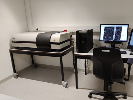
Bruker SkyScan 1172
The Skyscan 1172 is a microfocus desktop X-ray microtomograph, optimized for small samples and a plug-and-play scanning experience.
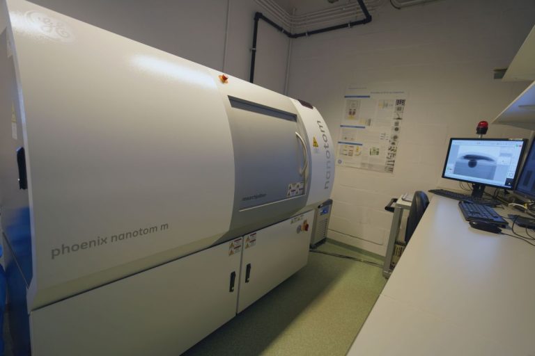
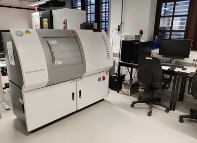
GE Nanotom M
The Nanotom M with its high-power nanofocus tube and stabilised ultra-precision mechanics invades the sub-micron range in voxel size.
GE Nanotom S
The Nanotom S with its high-power nanofocus tube and stabilised ultra-precision mechanics invades the sub-micron range in voxel size.
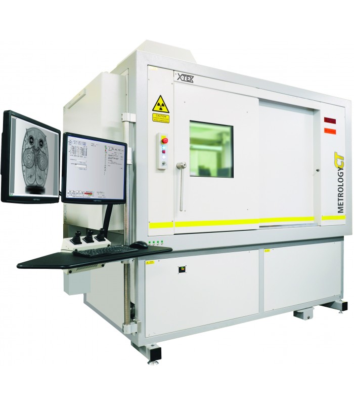
Nikon XT H 225 ST
The 225 kV system offers high power combined with high measurement accuracy suitable to a vast range of materials and sample sizes.
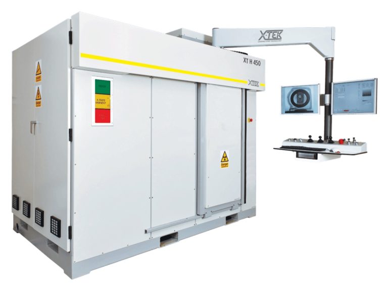
Nikon XT H 450
The 450 kV device (unique in Belgium) offers precise dimensional metrology and quality control for very large and dense components.

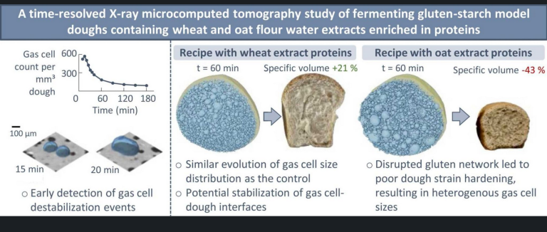
A time-resolved X-ray microcomputed tomography study of fermenting gluten-starch model doughs containing wheat and oat flour water extracts enriched in proteins
A B S T R A C T Water-extractable (WE) cereal flour constituents significantly influence bread loaf volume. However, the underlying mechanisms and the contribution
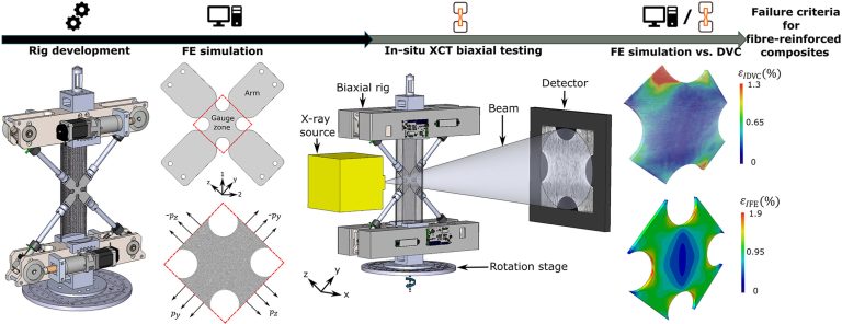
In situ biaxial tensile testing of composites: coupling X-ray computed tomography and digital volume correlation with finite element simulations
Abstract The mechanical behaviour of fibre-reinforced composites under multiaxial loading is critical for their structural performance but remains challenging to characterise at the microscale. This
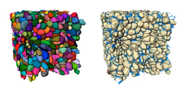
Panoptic segmentation for complete labeling of fruit microstructure in 3D micro-CT images with deep learning
Abstract Metabolic processes in plant organs involving transport of water, metabolic gasses, and nutrients depend on the three-dimensional (3D) microscopic tissue morphology. However, imaging and
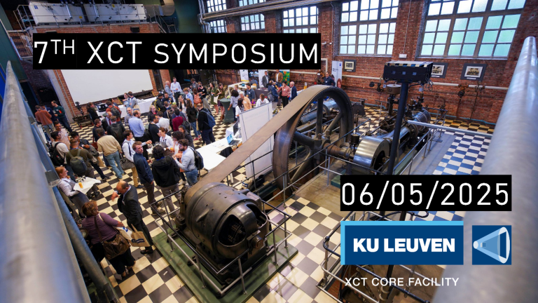
7th Symposium on X-ray Computed Tomography organized by the KU Leuven XCT Core Facility, to take place on May 6, 2025
Dear all, We are excited to announce the 7th Symposium on X-ray Computed Tomography organized by the KU Leuven XCT Core Facility, to take
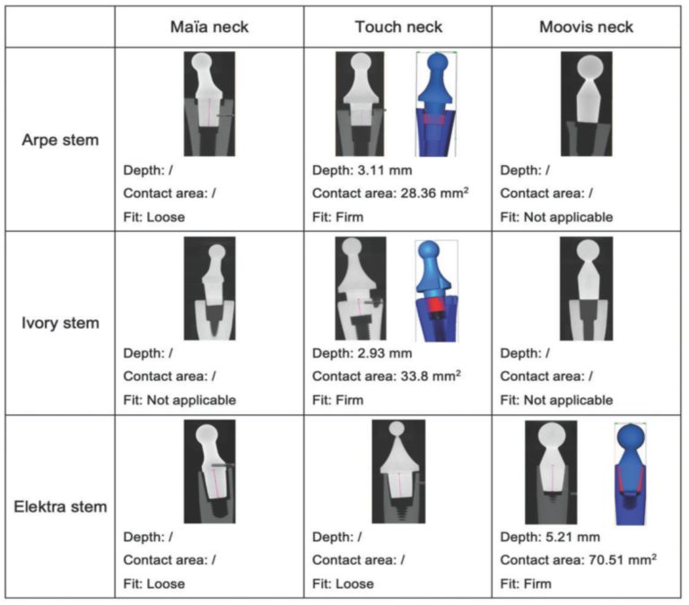
Revision options for discontinued trapeziometacarpal arthroplasties: compatibility with currently available implants
Abstract Surgical revision options for failed trapeziometacarpal total joint replacement include implant replacement and trapeziectomy. However, discontinuation of older implants complicates revision with original components,
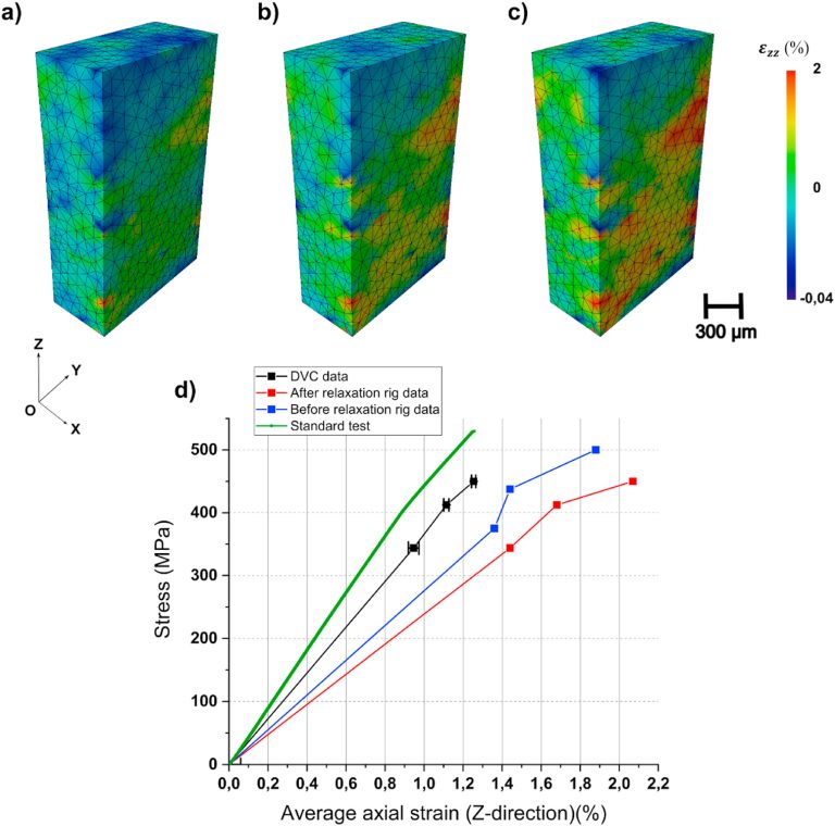
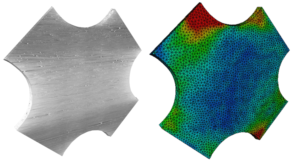
Spatial strain distribution and in-situ damage analysis of sheet moulding compounds based on digital volume correlation
Abstract Carbon fibre-reinforced thermoplastics sheet moulding compounds demonstrate significant potential for cost-effective, mass production applications in lightweight structures. However, the material’s complex internal morphology poses